• Laser & Optoelectronics Progress
  • Vol. 60, Issue 19, 1914001 (2023)
Shuaikang Cheng, Di Wu, Guohu Luo, and Yongxiang Hu*
Author Affiliations
  • State Key Laboratory of Mechanical System and Vibration, School of Mechanical Engineering, Shanghai Jiao Tong University, Shanghai 200240, China
  • show less
    DOI: 10.3788/LOP223006 Cite this Article Set citation alerts
    Shuaikang Cheng, Di Wu, Guohu Luo, Yongxiang Hu. Effect of Pulse Duration on Laser-Induced Forward Transfer of Microdroplets[J]. Laser & Optoelectronics Progress, 2023, 60(19): 1914001 Copy Citation Text show less
    Schematic of LIFT (a) process and (b) experimental setup
    Fig. 1. Schematic of LIFT (a) process and (b) experimental setup
    Physical pictures of experimental (a) setup and (b) samples
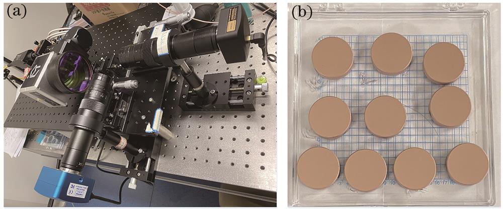
    Fig. 2. Physical pictures of experimental (a) setup and (b) samples
    SEM images of donor copper film and receiving substrate under different pulse durations. (a) 14 ns; (b) 20 ns; (c) 30 ns; (d) 50 ns; (e) 100 ns
    Fig. 3. SEM images of donor copper film and receiving substrate under different pulse durations. (a) 14 ns; (b) 20 ns; (c) 30 ns; (d) 50 ns; (e) 100 ns
    Processing map of LIFT under different pulse durations
    Fig. 4. Processing map of LIFT under different pulse durations
    SEM images of deposition microdroplets under different laser energy flux densities at pulse durations of 14 ns and 50 ns (sample rotated 55°). (a) 14 ns; (b) 50 ns
    Fig. 5. SEM images of deposition microdroplets under different laser energy flux densities at pulse durations of 14 ns and 50 ns (sample rotated 55°). (a) 14 ns; (b) 50 ns
    Schematic of formation of deposition microdroplet morphology under short pulse duration. (a) Single droplet; (b) stacked droplet; (c) fused droplet
    Fig. 6. Schematic of formation of deposition microdroplet morphology under short pulse duration. (a) Single droplet; (b) stacked droplet; (c) fused droplet
    Images of copper film and deposition microdroplet ablated by 20 ns, 4916 mJ/cm2 laser. (a) SEM image of copper film; (b) SEM image of deposition microdroplet; (c) confocal microscopy image of deposition microdroplet
    Fig. 7. Images of copper film and deposition microdroplet ablated by 20 ns, 4916 mJ/cm2 laser. (a) SEM image of copper film; (b) SEM image of deposition microdroplet; (c) confocal microscopy image of deposition microdroplet
    Material transfer rates under different pulse durations
    Fig. 8. Material transfer rates under different pulse durations
    Large-scale microdroplet array. (a) SEM image of microdroplet array; (b) mesh matching diagram of deposition microdroplet and ablative pit; (c) landing errors of Δx and Δy
    Fig. 9. Large-scale microdroplet array. (a) SEM image of microdroplet array; (b) mesh matching diagram of deposition microdroplet and ablative pit; (c) landing errors of Δx and Δy
    No.Pulse duration /nsRepetition rate /kHzSpot size /μmSingle pulse energy /μJEnergy division
    11420198.65‒16.5010
    22020199.00‒19.3015
    33020198.84‒19.2021
    45020198.83‒22.5023
    510020198.92‒23.9021
    Table 1. Experimental scheme of LIFT
    Shuaikang Cheng, Di Wu, Guohu Luo, Yongxiang Hu. Effect of Pulse Duration on Laser-Induced Forward Transfer of Microdroplets[J]. Laser & Optoelectronics Progress, 2023, 60(19): 1914001
    Download Citation