• Chinese Optics Letters
  • Vol. 23, Issue 2, 021102 (2025)
Chen Liang1,2, Junwei Wu2, Hangbing Peng2, Lijun Deng1..., Yiqin Lin2, Zhongwen Cheng2,*, Lüming Zeng1,2,** and Xuanrong Ji2|Show fewer author(s)
Author Affiliations
  • 1Jiangxi Provincial Key Laboratory of Advanced Electronic Materials and Devices, Jiangxi Science and Technology Normal University, Nanchang 330038, China
  • 2State Key Laboratory of Precision Electronics Manufacturing Technology and Equipment, Guangdong University of Technology, Guangzhou 510006, China
  • show less
    DOI: 10.3788/COL202523.021102 Cite this Article Set citation alerts
    Chen Liang, Junwei Wu, Hangbing Peng, Lijun Deng, Yiqin Lin, Zhongwen Cheng, Lüming Zeng, Xuanrong Ji, "In vivo whole brain photoacoustic microscopy through a transparent ultrasound transducer," Chin. Opt. Lett. 23, 021102 (2025) Copy Citation Text show less
    (a) Schematics of the TUT-based PAM system. L1, L2, and L3, lenses; GS, galvanometer scanner; TUT, transparent ultrasonic transducer; DAQ, data acquisition system; PC, personal computer; FPGA, field programmable gate array. (b) The PA image of the leaf venation skeleton. (c) Optical photo and PA image of a tilted tungsten filament inserted into an agar phantom. (d) The optical photo of the TUT. (e) The PA image of the whole mouse cortex vasculature.
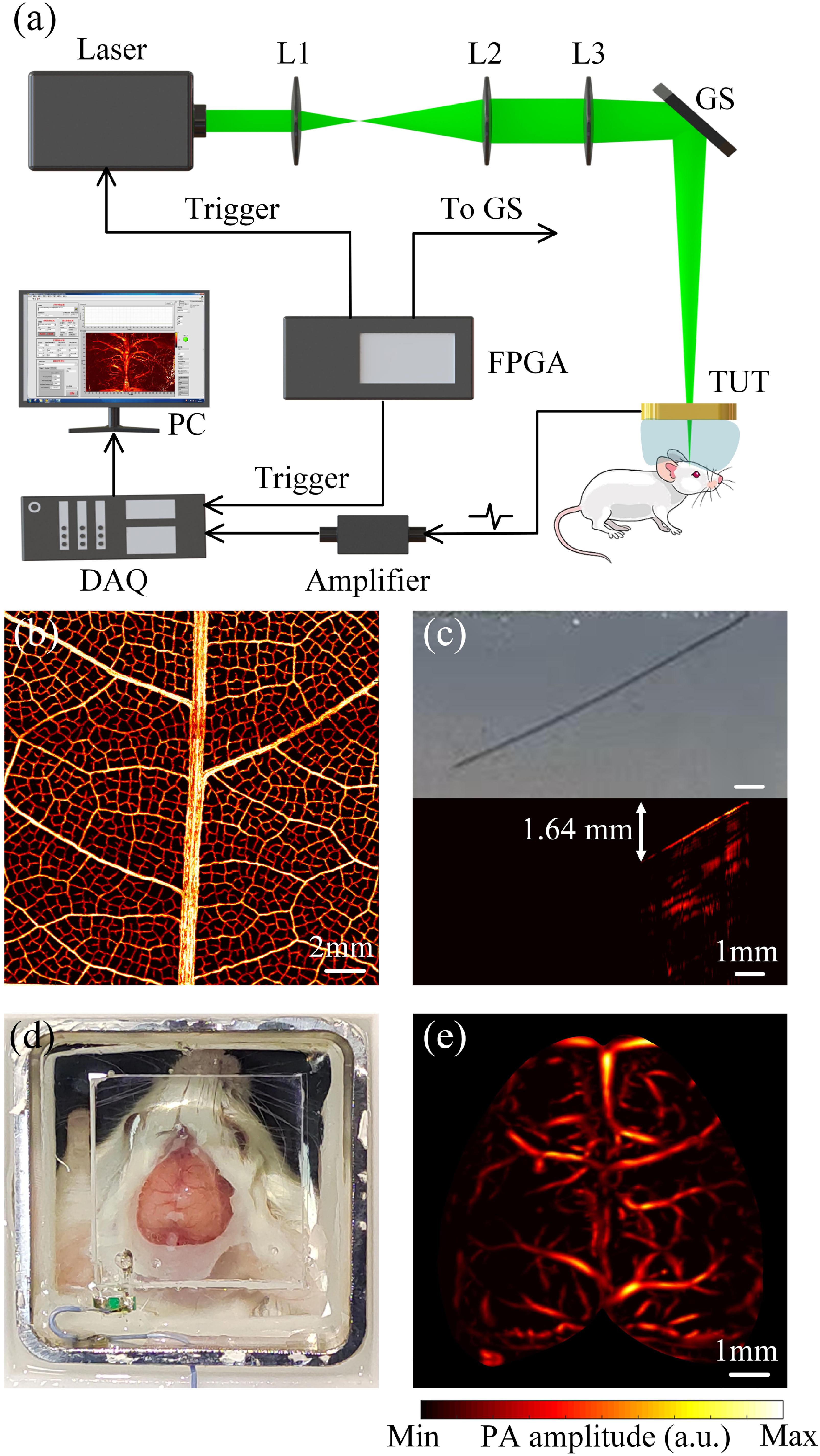
    Fig. 1. (a) Schematics of the TUT-based PAM system. L1, L2, and L3, lenses; GS, galvanometer scanner; TUT, transparent ultrasonic transducer; DAQ, data acquisition system; PC, personal computer; FPGA, field programmable gate array. (b) The PA image of the leaf venation skeleton. (c) Optical photo and PA image of a tilted tungsten filament inserted into an agar phantom. (d) The optical photo of the TUT. (e) The PA image of the whole mouse cortex vasculature.
    (a) The optical photo of a mouse before brain injury. (b) The PA image of the mouse before brain injury. (c) The optical photo of a mouse after brain injury. (d) The PA image of the mouse after brain injury. (e) Profiles of blood vessels traced along the white dashed lines in (b) and (d).
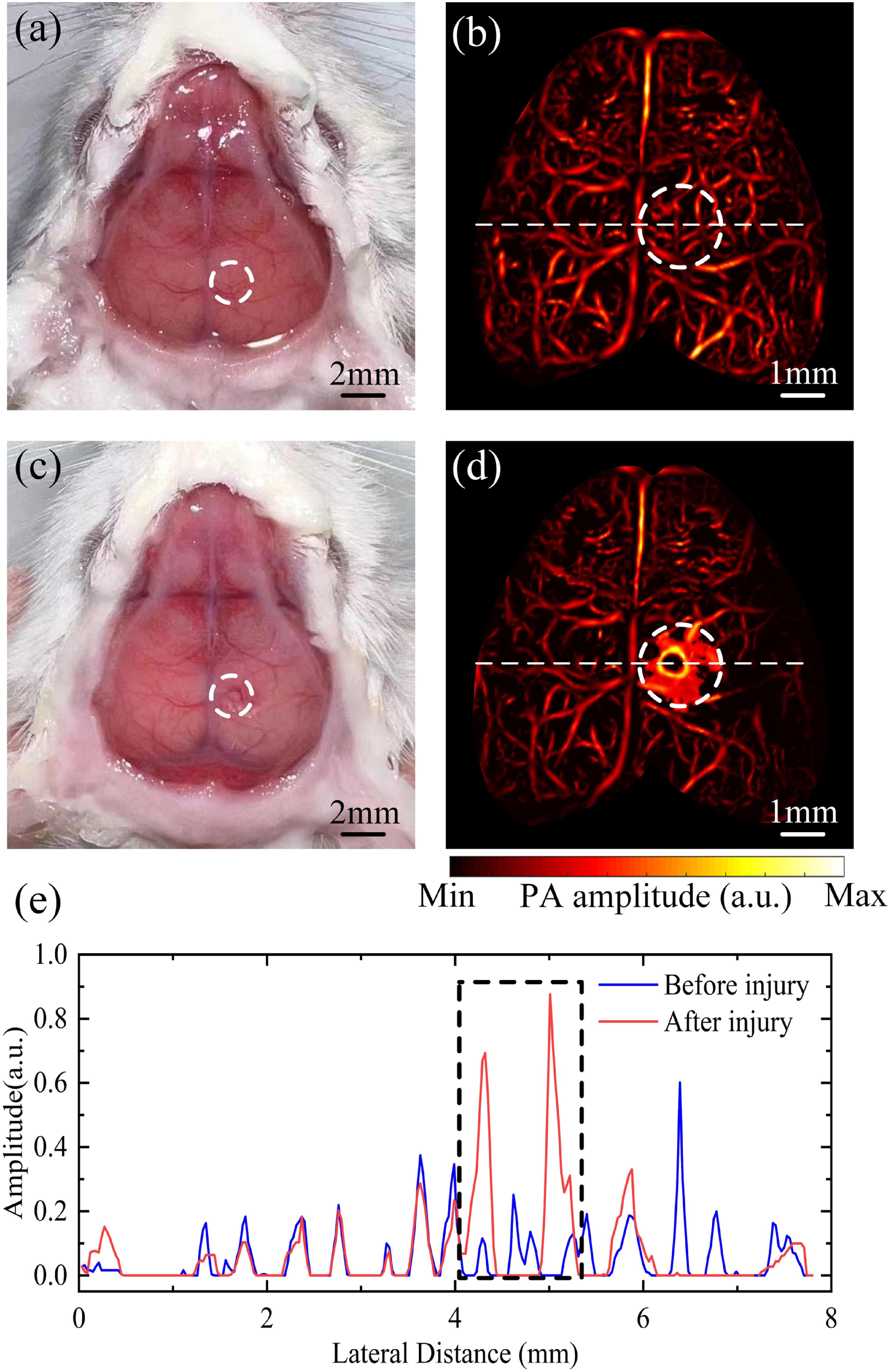
    Fig. 2. (a) The optical photo of a mouse before brain injury. (b) The PA image of the mouse before brain injury. (c) The optical photo of a mouse after brain injury. (d) The PA image of the mouse after brain injury. (e) Profiles of blood vessels traced along the white dashed lines in (b) and (d).
    (a) The optical photo of glioma in a nude mouse. (b) The PA image of the glioma. (c) The MRI imaging result of the glioma. (d) The HE staining result of the glioma.
    Fig. 3. (a) The optical photo of glioma in a nude mouse. (b) The PA image of the glioma. (c) The MRI imaging result of the glioma. (d) The HE staining result of the glioma.
    (a) Plot of changes in the cerebral cortex of a mouse 6 min and 40 s after hemorrhage. The location of the hemorrhage is circled by dotted lines. (b) Pixels and amplitudes in the white dashed circle over time.
    Fig. 4. (a) Plot of changes in the cerebral cortex of a mouse 6 min and 40 s after hemorrhage. The location of the hemorrhage is circled by dotted lines. (b) Pixels and amplitudes in the white dashed circle over time.
    Chen Liang, Junwei Wu, Hangbing Peng, Lijun Deng, Yiqin Lin, Zhongwen Cheng, Lüming Zeng, Xuanrong Ji, "In vivo whole brain photoacoustic microscopy through a transparent ultrasound transducer," Chin. Opt. Lett. 23, 021102 (2025)
    Download Citation