
Ziqi Li, Xiaolan Zhong, Chaohao Chen, Fan Wang. A New Weapon For Super-Resolution Imaging: Lanthanide Ion Doped Upconversion Nanoparticles (Invited)[J]. Laser & Optoelectronics Progress, 2024, 61(6): 0618018
- Laser & Optoelectronics Progress
- Vol. 61, Issue 6, 0618018 (2024)
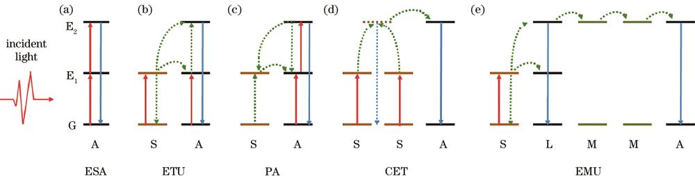
Fig. 1. Principal upconversion mechanisms of lanthanide-doped nanomaterials. (a) Excited-state absorption; (b) energy-transfer upconversion; (c) photon avalanche; (d) cooperative energy transfer; (e) energy migration-mediated upconversion (red arrows stand for direct excitation processes, blue arrows represent radiative emission processes, dashed arrows represent energy transfer processes, A means activator ion, S means sensitizer ion, M means migratory ion, and L means ladder ion)
![Principle of super-resolution microscopes. (a) Stimulated emission depletion microscope[50]; (b) fluorescence emission difference microscope[50]; (c) super-linear excitation-emission microscope[54]; (d) structure illumination microscopy[54]](/richHtml/lop/2024/61/6/0618018/img_02.jpg)
Fig. 2. Principle of super-resolution microscopes. (a) Stimulated emission depletion microscope[50]; (b) fluorescence emission difference microscope[50]; (c) super-linear excitation-emission microscope[54]; (d) structure illumination microscopy[54]
Fig. 3. Applications of UCNP in various super-resolution microscopy. (a) Super-resolution of low-power STED microscopy[21]; (b) immunofluorescence labeling of cellular cytoskeleton protein desmin with antibody-conjugated UCNPs and super-resolution imaging[45]; (c) non-bleached FED microscopy[48]; (d) Fourier-domain heterochromatic fusion[50]; (e) deep tissue imaging by near-infrared emission saturation microscopy[49]; (f) super-linear excitation-emission conventional confocal for three-dimensional subdiffraction imaging[52]; (g) super-resolution uSEE-STED imaging of UCNPs endocytosed by a neuron phenotype cell[53]; (h) nonlinear SIM microscopy super-resolution[56]
Fig. 4. Applications of UCNP in Multiplexed super-resolution microscopy. (a) Deep learning assisted decoding of multi-channel single τ2-dots in super-resolution mode[57]; (b) super-resolved multiplexing of mixed UCNPs[58]; (c) time-domain anticounterfeiting by using three types of Nd-Yb-Tm series τ2-dots security inks with different rising-decay fingerprints; (d) multiplexed single molecule digital assays using five types of Nd-Yb-Er series τ2-dots probes to quantify the five kinds of target pathogen single-stranded DNAs[13]


Set citation alerts for the article
Please enter your email address

© Copyright 2018-2021 | Chinese Laser Press. All Rights Reserved 沪ICP备15018463号-20