• Laser & Optoelectronics Progress
  • Vol. 60, Issue 10, 1000002 (2023)
Xiupin Wu1,*, Juewei Li1, and Wanrong Gao2
Author Affiliations
  • 1School of Medical Instruments, Shanghai University of Medicine & Health Sciences, Shanghai 201318, China
  • 2School of Electronic and Optical Engineering, Nanjing University of Science & Technology, Nanjing 210014, Jiangsu, China
  • show less
    DOI: 10.3788/LOP220794 Cite this Article Set citation alerts
    Xiupin Wu, Juewei Li, Wanrong Gao. Public Data Acquisition of Optical Coherence Tomography Images of Fundus and Its Analysis Algorithms[J]. Laser & Optoelectronics Progress, 2023, 60(10): 1000002 Copy Citation Text show less
    Macular structure of healthy retina
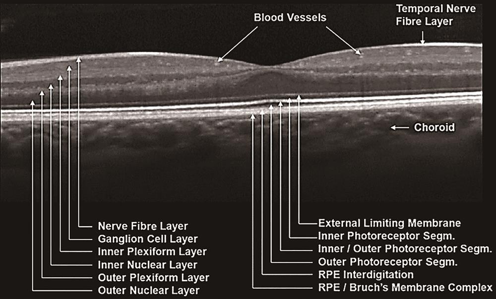
    Fig. 1. Macular structure of healthy retina
    OCT image of AMD
    Fig. 2. OCT image of AMD
    OCT image of DR
    Fig. 3. OCT image of DR
    OCT image of CSR
    Fig. 4. OCT image of CSR
    Number of papers of different algorithms
    Fig. 5. Number of papers of different algorithms
    DatasetDevicesEye diseaseNumber of imagesCountryLink to datasetYear
    DukeHome-madeAMD,Normal>38400USAhttps://people.duke.edu/~sf59/RPEDC_Ophth_2013_dataset.htm2013—2021
    KaggleAMD,CNV,CSR,DME,DR,Drusen,MH,Normal>3000Melbournehttps://www.kaggle.com/search?q=Retinal+OCT+in%3Adatasets2017—2021
    VarpaCirrus 500,Cal Zeiss353Spainhttp://www.varpa.es/research/ophtalmology.html2010—2019
    OCTIDCirrus 500,Cal ZeissDR,AMD,CSR,Normal519Canadahttps://dataverse.scholarsportal.info/dataverse/OCTID2018
    ZenodoCirrus 500,Cal ZeissGlaucoma,Normal1100https://zenodo.org/record/1481223#.Xr06Q2gzbIU2018
    MendeleySpectralis,HeidelbergDR,Drusen,CNV,Normal84484USA,Chinahttps://data.mendeley.com/datasets/rscbjbr9sj/32013—2017
    OCTRIMASpectralis,HeidelbergNormal10USAhttps://journals.plos.org/plosone/article?id=10.1371/journal.pone.01339082015
    Pascal DufourSpectralis,HeidelbergAMD,Normal310Switzerlandhttp://pascaldufour.net/Research/software_data.html2012
    Table 1. Free public data
    CompanyModelCentral wavelengthResolutionField of viewLine ratePixel number
    Cal Zeiss,USCirrus 500 OCT840 nm5 μm×15 μm2 mm27-68 kHz1024
    Topcon,JapanDRI OCT Triton,1050 nm8 μm×20 μm100 kHz
    3D OCT-1 Maestro,3D OCT-20006 μm×20 μm50 kHz
    Heidelberg,GermanySpectralis1 μm40 kHz
    Optovue,USRTVue-XR5 μm2.3 mm70 kHz
    Bioptigen,USSD-OCT<3 μm5.4 mm32 kHz1024/2048
    Leica,GermanyEnvisu SD-OCT≤2μm
    Table 2. OCT devices for data acquisition
    TypeMethodEye diseaseDatasetResearch instituteYear
    Different layersU-net and SegNet15AMD,DMEDukeTowson University,USA2021
    U-net and DexiNet16AMDDukeUniversity of Florida,USA2021
    GS17DME,AMD,NormalOCTRIMAUniversity of Miami,USA2015
    GS18AMDDukeDoheny Eye Institute,USA2017
    ERR-net and GS19AMD,DRDukeXiangtan University,China2021
    CNN20AMD,NormalDukeUniversity of Iowa,USA2020
    CNN and LSTM21AMD,NormalDukeIndia2017
    CNN22AMDDukeUniversity of Iowa,USA2017
    CNN23AMD,NormalDukeUniversity of Iowa,USA2018
    CNN-GS24AMDDukeDuke University,USA2017
    GDM25AMD,NormalOCTRIMAUniversity of Nottingham,UK2017
    Fuzzy histogram and graph cut methods26GlaucomaDukeBrunel University,UK2018
    Based on fluid potential energy equation27AMDDukeTianjin University,China2020
    MT and probabilistic maps28AMDDukeIndia2018
    Bayesian deep learning29DME,AMDDukeAustralia2018
    Non-parametric mathematical model30AMD,NormalDukeIsfahan University of Medical Sciences,Iran2021
    CRF31DME,AMD,NormalOCTRIMA,DukeInternational Institute of Information Technology Hyderabad,India2018
    Ensemble-learning-based method32DME,NormalDukeUniversity of Coimbra,Portugle2021
    Structured learning algorithm33DMEPascal DufourIndia2016
    Lesion zoneU-net34AMDDukeAustria2021
    U-net++35DMEDukeHeilongjiang University of Science and Technology,China2020
    Multitask learning36AMDDukeMedical University of Vienna,Austria2019
    CycleGAN-based weakly supervised method37DME,CNV,Drusen,NormalMendeleySuzhou Institute of Biomedical Engineering and Technology,China2021
    Structure tensor guided approach38AMD,CSR,DR,CNV,MH,NormalDuke,MendeleyBeihang University,China2021
    RASP-Net39AMDMendeleyBeihang University,China2021
    Unsupervised learning40AMDDukeMedical University Vienna,Austria2019
    Gradient-weighted class activation mapping41DME,CNV,Drusen,NormalMendeleyWuhan University of Science and Technology,China2022
    Encoder-decoder network42DR,NormalVerpaShenzhen University,China2021
    BSDA-Net43DR,NormalVerpaSouthern University of Science and Technology,China2021
    Table 3. Segmentation algorithm for retinal OCT images
    MethodEye diseaseDatasetAccuracyResearch instituteYear
    CNN44AMD,DR,NormalPascal Dufour88%University of Bern,Switzerland2019
    CNN45AMD,DME,NormalDukeAUC is 0.95Indian Institute of Technology Guwahati,India2020
    CNN46AMD,DME,NormalMendeley95.7%Sri Sivasubramaniya Nadar College of Engineering,India2021
    CNN47AMD,DME,NormalDukeIndia2017
    Multiscale and multipath CNN48AMDDuke,Mendeley99.02%National Institute of Technology Tiruchirappalli,India2021
    Lesion-ware CNN49Drusen,CNV,DME,NormalMendeley93.3%Hunan University,China2019
    Self-developed CNN50CNV,DME,Drusen,NormalMendeley94.87%Bangladesh2021
    Three CNNs51CNV,DME,Drusen,NormalKaggle96.5%India2022
    Densely connected CNN52CNV,DME,DrusenMendeley98.0%National Institute of Technology Rourkela,India2019
    CNN and quadratic SVM53AMD,CNV,DME,Drusen,NormalMendeley100%University of Southern Queensland,Australia2021
    SVM54AMD,NormalDuke95%National University of Sciences and Technology,Pakistan2018
    Multilayered SVM55AMD,CSR,NormalDuke99.92%Bahria University,Pakistan2017
    GAN56CNV,DME,Drusen,NormalMendeley93.9%Aerospace Medical Center,Republic of Korea2021
    LSGAN57CNV,DME,Drusen,NormalMendeley87.25%Hunan University,China2020
    VGG1658CNV,DME,Drusen,NormalMendeley98.7%National Institutes of Health,USA2021
    Deep ensemble network59CNV,DME,Drusen,NormalMendeley98.53%India2020
    ResNet60AMDDuke96.78%Institute for Infocomm Research,Singapore2020
    GLMR61AMD,NormalDukeAUC>0.99Duke University,USA2014
    SMO62AMD,DME,NormalDuke99.3%Northeastern University,China2016
    CMoE63AMD,DME,NormalDuke98.14%Isfahan University of Medical Sciences,Iran2019
    MCME ensemble model64AMD,DME,NormalDuke98.86%Isfahan University of Medical Sciences,Iran2018
    Super-learner framework65CNV,DME,Drusen,NormalMendeley98.55%India2021
    Vision Transformer66AMD,DME,NormalDuke99.69%Shanghai Institute of Technology,China2022
    Multiscale CNN67AMD,NormalOCTID,Duke>96.0%National Institute of Technology Tiruchirappalli,India2021
    Table 4. Classification algorithm of retinal OCT images
    MethodDatasetMetricCountryYear
    AC-SRResNet68MendeleySNRCNREPISpeedBeijing Institute of Technology,China2021
    41.8 dB42.0 dB0.6817 frame/s
    2D curvelet-based K-SVD71DukeCNRMSRNoor Eye Hospital,Iran2018
    5.1214.41
    NWSR72DukeMSRCNRENLPSNRUniversity of Isfahan,Iran2018
    8.673.451552.6827.79
    GAN69DukePSNREPIENLCNRSichuan University,China2019
    27.830.931669.54.46
    Table 5. Denoising algorithm of retinal OCT images
    Xiupin Wu, Juewei Li, Wanrong Gao. Public Data Acquisition of Optical Coherence Tomography Images of Fundus and Its Analysis Algorithms[J]. Laser & Optoelectronics Progress, 2023, 60(10): 1000002
    Download Citation