
Yingzhe Gao, Yi Yuan, Zhenhe Ma. High-Resolution Cortical Blood Flow Imaging Based on Optical Coherence Tomography[J]. Laser & Optoelectronics Progress, 2019, 56(11): 111101
- Laser & Optoelectronics Progress
- Vol. 56, Issue 11, 111101 (2019)
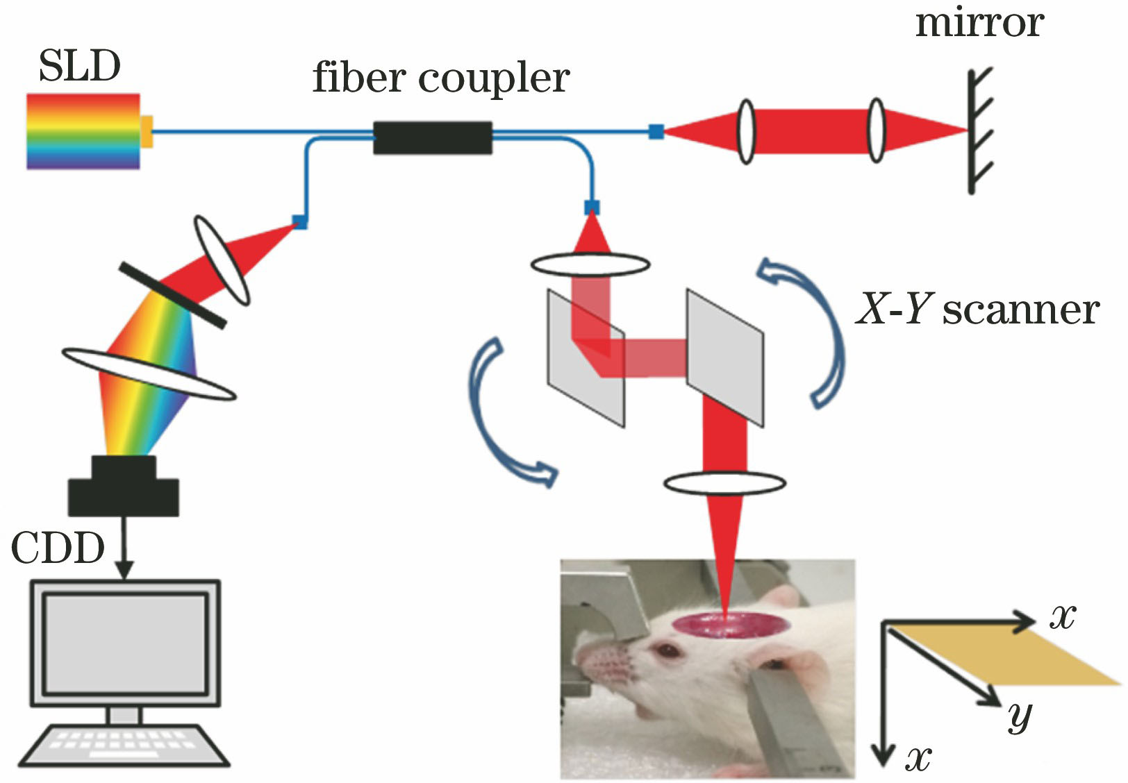
Fig. 1. Schematic of spectral OCT angiography system

Fig. 2. Lateral resolution measurement with knife edge method
Fig. 3. Dispersion compensation for axial resolution maintaining
Fig. 4. High-resolution imaging results by spectral OCT angiography system. (a) Image of two-dimensional structure by OCT; (b) image of flow signal by OCT with ① and ② indicating dural blood vessels and large blood vessels on cerebral cortex, respectively; (c) X-Y projection display of OCT vascular image; (d) three-dimensional display of OCT vascular images; (e) cerebral dural vascular image; (f) cerebral cortical vascular image
Fig. 5. Whole cerebral vascular image with rat brain photograph shown in upper-left corner


Set citation alerts for the article
Please enter your email address

© Copyright 2018-2021 | Chinese Laser Press. All Rights Reserved 沪ICP备15018463号-20