• Laser & Optoelectronics Progress
  • Vol. 61, Issue 6, 0618005 (2024)
Fangrui Lin, Yiqiang Wang, Min Yi, Chenshuang Zhang..., Liwei Liu and Junle Qu*|Show fewer author(s)
Author Affiliations
  • Key Laboratory of Optoelectronic Devices and Systems of Ministry of Education/Guangdong Province, College of Physics and Optoelectronic Engineering, Shenzhen University, Shenzhen 518060, Guangdong, China
  • show less
    DOI: 10.3788/LOP240467 Cite this Article Set citation alerts
    Fangrui Lin, Yiqiang Wang, Min Yi, Chenshuang Zhang, Liwei Liu, Junle Qu. Research Progress on Fast Fluorescence Lifetime Imaging Microscopy and Its in vivo Applications (Invited)[J]. Laser & Optoelectronics Progress, 2024, 61(6): 0618005 Copy Citation Text show less
    Number of papers focusing on FLIMfor in vivo imaging
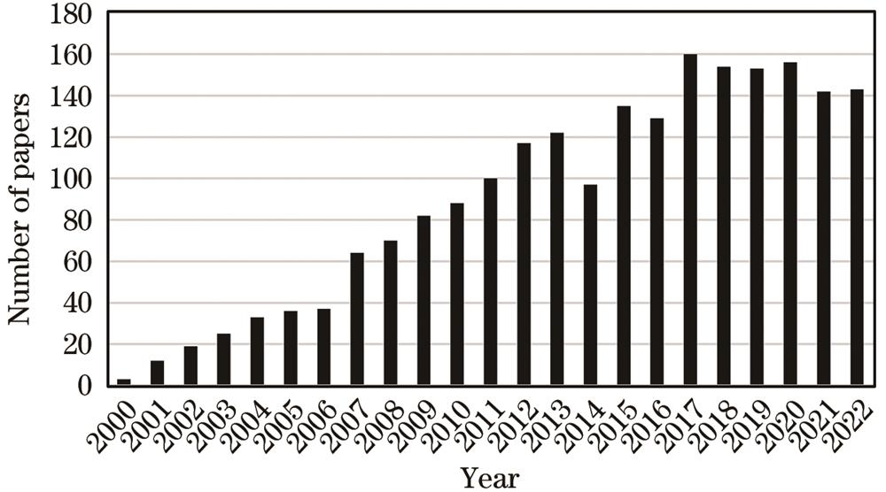
    Fig. 1. Number of papers focusing on FLIMfor in vivo imaging
    Principle diagram of TD-FLIM and FD-FLIM
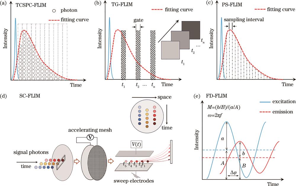
    Fig. 2. Principle diagram of TD-FLIM and FD-FLIM
    Imaging speed improving of TCSPC-FLIM by optimizing detection methods. (a) Using a collection module with a shorter dead time to record multiple photons within a single pulse cycle[36]; (b) using a photon'Spinner' to shorten the dead time of the capture card[37]; (c) using parallel array detection modules to improve photon counting rate[38]
    Fig. 3. Imaging speed improving of TCSPC-FLIM by optimizing detection methods. (a) Using a collection module with a shorter dead time to record multiple photons within a single pulse cycle[36]; (b) using a photon'Spinner' to shorten the dead time of the capture card[37]; (c) using parallel array detection modules to improve photon counting rate[38]
    Imaging speed improving of TCSPC-FLIM by optimizing scanning methods. (a) Implementing fast scanning with arbitrary shapes based on 2D-AOD[39]; (b) using a multifocal array to achieve fast scanning[41]; (c) using line excitation to realize fast scanning, and the imaging results of two slices shown on right[44]
    Fig. 4. Imaging speed improving of TCSPC-FLIM by optimizing scanning methods. (a) Implementing fast scanning with arbitrary shapes based on 2D-AOD[39]; (b) using a multifocal array to achieve fast scanning[41]; (c) using line excitation to realize fast scanning, and the imaging results of two slices shown on right[44]
    Schematic of TG-FLIM system based on light-sheet[55]. (a) Schematic of the system; (b) imaging results of a living zebrafish
    Fig. 5. Schematic of TG-FLIM system based on light-sheet[55]. (a) Schematic of the system; (b) imaging results of a living zebrafish
    SC-FLIM technique for rapid imaging. (a) Single particle tracking FLIM based on double helix point spread function engineering[67]; (b) implementation of high-speed FLIM using compressed sensing-based DMD spatial encoding technology[68]
    Fig. 6. SC-FLIM technique for rapid imaging. (a) Single particle tracking FLIM based on double helix point spread function engineering[67]; (b) implementation of high-speed FLIM using compressed sensing-based DMD spatial encoding technology[68]
    Widefield FD-FLIM based on modulation of LED[70]. (a) Schematic of the system; (b) fluorescence intensity images of Fluorescein-Glycerol (Fl-Gly) solutions with different ratios; (c) corresponding fluorescence lifetime images
    Fig. 7. Widefield FD-FLIM based on modulation of LED[70]. (a) Schematic of the system; (b) fluorescence intensity images of Fluorescein-Glycerol (Fl-Gly) solutions with different ratios; (c) corresponding fluorescence lifetime images
    Correcting motion artifacts for in vivo FLIM through post-processing. (a) Inter frame artifact correction based on normalized cross-correlation algorithm[77]; (b) inter frame and intra frame artifact correction based on the Lucas-Kanade framework[78]
    Fig. 8. Correcting motion artifacts for in vivo FLIM through post-processing. (a) Inter frame artifact correction based on normalized cross-correlation algorithm[77]; (b) inter frame and intra frame artifact correction based on the Lucas-Kanade framework[78]
    Flowchart of ADCG algorithm and the fitting results with photon number of 45[86]
    Fig. 9. Flowchart of ADCG algorithm and the fitting results with photon number of 45[86]
    Three types of DL algorithms contribute to reduce FLIM processing time. (a) Traditional curve fitting is substituted by either a 3D-CNN[92] (left) or a 1D-CNN[93] (right) framework; (b) LLE and NIII sub networks are utilized to produce data with high photon counts from low photon counts[96]; (c) DL algorithms are employed to improve imaging resolution[97]
    Fig. 10. Three types of DL algorithms contribute to reduce FLIM processing time. (a) Traditional curve fitting is substituted by either a 3D-CNN[92] (left) or a 1D-CNN[93] (right) framework; (b) LLE and NIII sub networks are utilized to produce data with high photon counts from low photon counts[96]; (c) DL algorithms are employed to improve imaging resolution[97]
    Schematic of instant FLIM technique and its in vivo imaging results of neurons in mouse brain[71]
    Fig. 11. Schematic of instant FLIM technique and its in vivo imaging results of neurons in mouse brain[71]
    TCSPC-FLIM for living mice. (a) Imaging brain vessels and Aβ plaque in AD mice[103]; (b) quantitative characterization of intracellular glucose concentration in cortical neurons[104]; (c) three photon FLIM of cerebral vessels[105]; (d) imaging of different organs in mice based on microendoscopy[106]
    Fig. 12. TCSPC-FLIM for living mice. (a) Imaging brain vessels and Aβ plaque in AD mice[103]; (b) quantitative characterization of intracellular glucose concentration in cortical neurons[104]; (c) three photon FLIM of cerebral vessels[105]; (d) imaging of different organs in mice based on microendoscopy[106]
    Applications of FLIM for intraoperative tumor diagnosis. (a) Recognition of tumor boundary based on PS-FLIM[108]; (b) high-resolution recognition of tumor cells based on TCSPC-FLIM[115]; (c) fast FD-FLIM with a large field-of-view based on 5-ALA labeling[117]
    Fig. 13. Applications of FLIM for intraoperative tumor diagnosis. (a) Recognition of tumor boundary based on PS-FLIM[108]; (b) high-resolution recognition of tumor cells based on TCSPC-FLIM[115]; (c) fast FD-FLIM with a large field-of-view based on 5-ALA labeling[117]
    Network structureTraining timePrediction timePhoton count
    ANN904 h0.9 s(256×256 pixel)<900
    CNN914.5 h<3 ms(32×32 pixel)25‒1600
    3D CNN92~2.5 s(512×512 pixel)250 or 500
    1D CNN93a few minutesa few seconds(256×256 pixel)
    GAN94

    GAN:6.1 h

    Estimator:0.1 h

    ~0.15‒0.17 ms50‒200
    LLE and NIII960.63 s(256×256 pixel)4‒10
    τ-Net and spatial resolution improved net97400 ha few seconds(256×256 pixel)100‒10000
    Table 1. Performance comparison of several fast FLIM technologies based on DL
    Fangrui Lin, Yiqiang Wang, Min Yi, Chenshuang Zhang, Liwei Liu, Junle Qu. Research Progress on Fast Fluorescence Lifetime Imaging Microscopy and Its in vivo Applications (Invited)[J]. Laser & Optoelectronics Progress, 2024, 61(6): 0618005
    Download Citation