
Keyi Fei, Bingying Lin, Zhongzhou Luo, Yupei Chen, Jin Yuan, Peng Xiao, "Label-free cellular imaging of mouse retina with dual-mode full-field optical coherence tomography," Chin. Opt. Lett. 23, 031701 (2025)
- Chinese Optics Letters
- Vol. 23, Issue 3, 031701 (2025)
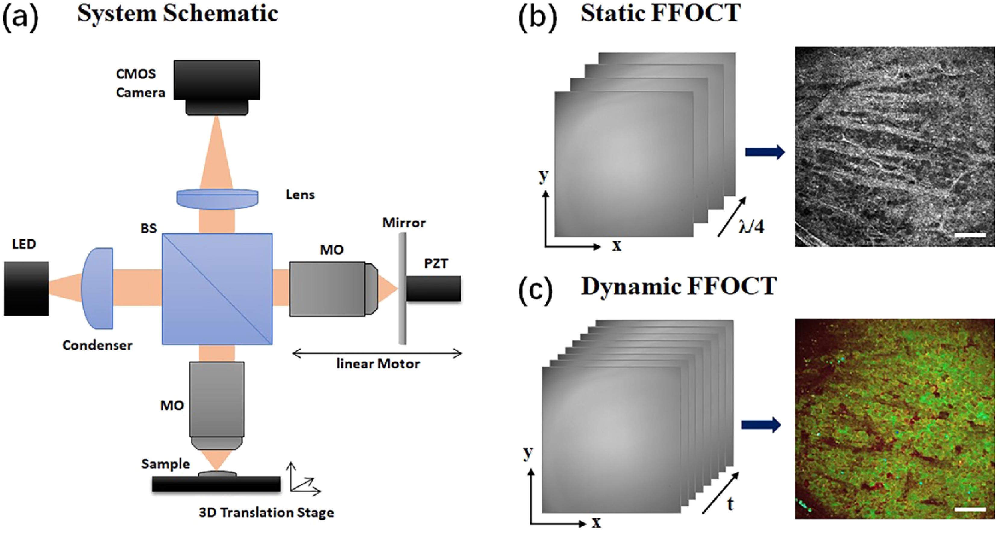
Fig. 1. (a) The optical schematic of our customized dual-mode FFOCT system, (b) static FFOCT image acquired with a 4-phase modulation scheme, and (c) D-FFOCT image extracted through power spectral density analysis color-coding of temporal FFOCT interferograms. BS: beam splitter; LED: light emitting diode; MO: microscope objective; PZT: piezoelectric transducer. Scale bar: 50 µm.
![The depth-resolved static (upper) and dynamic (lower) FFOCT images of normal mouse retina acquired with the dual-mode FFOCT system covering the (a), (b) nerve fiber layer, (c), (d) ganglion cell layer, (e), (f) inner plexiform layer, inner nuclear layer [(g), (h) upper layer; (i), (j) lower layer], outer plexiform layer [upper right corner of (i), (j)], and (k), (l) outer nuclear layer. Scale bar: 50 µm.](/richHtml/col/2025/23/3/031701/img_002.jpg)
Fig. 2. The depth-resolved static (upper) and dynamic (lower) FFOCT images of normal mouse retina acquired with the dual-mode FFOCT system covering the (a), (b) nerve fiber layer, (c), (d) ganglion cell layer, (e), (f) inner plexiform layer, inner nuclear layer [(g), (h) upper layer; (i), (j) lower layer], outer plexiform layer [upper right corner of (i), (j)], and (k), (l) outer nuclear layer. Scale bar: 50 µm.
Fig. 3. Dual-mode FFOCT images and the corresponding immunofluorescence images of the GCL acquired in early I/R injured mouse retinal tissue (upper) compared to normal mouse retinal tissue (lower). Scale bar: 50 µm.
Fig. 4. Dual-mode FFOCT image NFL in the nasal side and the vessels in the optic disk area. Scale bar: 50 µm.


Set citation alerts for the article
Please enter your email address

© Copyright 2018-2021 | Chinese Laser Press. All Rights Reserved 沪ICP备15018463号-20