• Journal of Radiation Research and Radiation Processing
  • Vol. 43, Issue 1, 010701 (2025)
Rui LUO*, Yuhe ZHANG, and Naqing MAO
Author Affiliations
  • National Key Laboratory for Metrology and Calibration Techniques, China Institute of Atomic Energy, Beijing 102413, China
  • show less
    DOI: 10.11889/j.1000-3436.2024-0045 Cite this Article
    Rui LUO, Yuhe ZHANG, Naqing MAO. Development of a method for measuring low energy electron beam irradiation parameters by calorimetry and step stacking[J]. Journal of Radiation Research and Radiation Processing, 2025, 43(1): 010701 Copy Citation Text show less
    Schematic diagram of low energy electron energy measurement
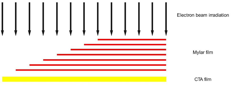
    Fig. 1. Schematic diagram of low energy electron energy measurement
    Calorimeter structure
    Fig. 2. Calorimeter structure
    Dose distribution of CTA film measurement with length, dotted line indicates the background baseline of the film dosimeter, red line indicates the number of Mylar film layers (color online)
    Fig. 3. Dose distribution of CTA film measurement with length, dotted line indicates the background baseline of the film dosimeter, red line indicates the number of Mylar film layers (color online)
    Simulation of energy deposition of electron beam in graphite material (density is 1.77 g/cm3). The generated electrons penetrate the 10 μm titanium window, 5 cm air, and Mylar film for complete energy deposition
    Fig. 4. Simulation of energy deposition of electron beam in graphite material (density is 1.77 g/cm3). The generated electrons penetrate the 10 μm titanium window, 5 cm air, and Mylar film for complete energy deposition
    Relationship between absorbed dose and reciprocal of the moving speed (a), and beam intensity (b)
    Fig. 5. Relationship between absorbed dose and reciprocal of the moving speed (a), and beam intensity (b)
    Comparison of specific heat value of graphite before and after irradiation
    Fig. 6. Comparison of specific heat value of graphite before and after irradiation
    Temperature change curve with time before and after irradiation
    Fig. 7. Temperature change curve with time before and after irradiation

    电子束能量 / keV

    Electron beam energy

    质量阻止本领 / (MeV·cm2·g-1)

    Mass stopping power

    CSDA射程 / (g·cm-2)

    CSDA range

    石墨吸收体中射程 / mm

    Range in graphite absorber

    1003.680.0160.073
    1303.210.0230.106
    1502.890.0320.143
    1802.660.0410.184
    2002.490.0500.228
    2502.250.0720.325
    3002.090.0950.430
    Table 1. Electron range for the energy from 100 keV to 300 keV

    测量器具

    Measuring instrument

    吸收剂量 / kGy

    Absorbed dose

    测量不确定度 / kGy

    Uncertainty

    量热计装置Calorimeter18.82.07
    丙氨酸剂量计Alanine dosimeter19.20.93
    Table 2. Comparison of measurement results between calorimeter and alanine dosimeter under the same condition

    不确定度来源

    Sources of uncertainty

    类型

    Type

    相对标准不确定度 / %

    Relative standard uncertainty

    特征比热容

    Heat capacity

    石墨成分 Graphite compositionB0.10
    石墨比热测量 Graphite specific heat measurementB0.64
    温度对比热影响 Effect of temperature on specific heatB0.37

    温升

    Temperature rising

    电阻特征曲线校准 Thermistor calibrationB0.52
    电阻测量 Thermistor measurementB0.02
    热损失修正 Heat loss correctionB4.43
    试验装置稳定性 The stabilityB1.12
    测量重复性 Measurement repeatabilityA1.86
    合成标准不确定度(uc)Combined standard uncertainty (uc)5.1
    扩展不确定度(U)(k=2)Extended uncertainty (U)(k=2)11
    Table 3. Uncertainty evaluation of 140 keV electron beam irradiation absorbed dose measurement
    Rui LUO, Yuhe ZHANG, Naqing MAO. Development of a method for measuring low energy electron beam irradiation parameters by calorimetry and step stacking[J]. Journal of Radiation Research and Radiation Processing, 2025, 43(1): 010701
    Download Citation