• Laser & Optoelectronics Progress
  • Vol. 55, Issue 3, 030006 (2018)
Lujia Jin, Yang He, Luxi Qu, Chi Zhang..., Meiqi Li* and Peng Xi|Show fewer author(s)
Author Affiliations
  • Department of Biomedical Engineering, College of Engineering, Peking University, Beijing 100871, China
  • show less
    DOI: 10.3788/LOP55.030006 Cite this Article Set citation alerts
    Lujia Jin, Yang He, Luxi Qu, Chi Zhang, Meiqi Li, Peng Xi. Analysis of New Super-Resolution Microscopy Technology[J]. Laser & Optoelectronics Progress, 2018, 55(3): 030006 Copy Citation Text show less
    References

    [1] Hao X, Kuang C F, Gu Z T et al. From microscopy to nanoscopy via visible light[J]. Light Science & Applications, 2, e108(2013). http://www.nature.com/lsa/journal/v2/n10/abs/lsa201364a.html

    [2] Yang X S, Xie H, Alonas E et al. Mirror-enhanced super-resolution microscopy[J]. Light Science & Applications, 5, e16134(2016). http://pubmedcentralcanada.ca/pmcc/articles/PMC4936537/

    [3] Hell S W. Microscopy and its focal switch[J]. Nature Methods, 6, 24-32(2009). http://old.med.wanfangdata.com.cn/viewHTMLEn/PeriodicalPaper_JJ025899021.aspx

    [4] Gustafsson M G L. Nonlinear structured-illumination microscopy: Wide-field fluorescence imaging with theoretically unlimited resolution[J]. Proceedings of the National Academy of Sciences of the United States of America, 102, 13081-13086(2005). http://bfg.oxfordjournals.org/cgi/ijlink?linkType=ABST&journalCode=pnas&resid=102/37/13081

    [5] Betzig E, Patterson G H, Sougrat R et al. Imaging intracellular fluorescent proteins at nanometer resolution[J]. Science, 313, 1642-1645(2006). http://jmicro.oxfordjournals.org/cgi/ijlink?linkType=ABST&journalCode=sci&resid=313/5793/1642

    [6] Hess S T. Girirajan T P K, Mason M D. Ultra-high resolution imaging by fluorescence photoactivation localization microscopy[J]. Biophysical Journal, 91, 4258-4272(2006).

    [7] Rust M J, Bates M, Zhuang X. Sub-diffraction-limit imaging by stochastic optical reconstruction microscopy (STORM)[J]. Nature Methods, 3, 793-795(2006). http://europepmc.org/abstract/MED/16896339

    [8] Dertinger T, Colyer R, Iyer G et al. Fast, background-free, 3D super-resolution optical fluctuation imaging (SOFI)[J]. Proceedings of the National Academy of Sciences, 106, 22287-22292(2009). http://jmicro.oxfordjournals.org/cgi/ijlink?linkType=ABST&journalCode=pnas&resid=106/52/22287

    [9] Yahiatene I, Hennig S, Müller M et al. Entropy-based super-resolution imaging (ESI): From disorder to fine detail[J]. ACS Photonics, 2, 1049-1056(2015). http://pubs.acs.org/doi/pdf/10.1021/acsphotonics.5b00307

    [10] Chen F, Tillberg P W, Boyden E S. Expansion microscopy[J]. Science, 347, 543-548(2015).

    [11] Marblestone A H, Boyden E S. Designing tools for assumption-proof brain mapping[J]. Neuron, 83, 1239-1241(2014). http://europepmc.org/articles/PMC4450254

    [12] Geertsema H, Ewers H. Expansion microscopy passes its first test[J]. Nature Methods, 13, 481-482(2016). http://www.nature.com/nmeth/journal/v13/n6/abs/nmeth.3872.html

    [13] Tillberg P W, Chen F, Piatkevich K D et al. Protein-retention expansion microscopy of cells and tissues labeled using standard fluorescent proteins and antibodies[J]. Nature Biotechnology, 34, 987-992(2016). http://www.nature.com/nbt/journal/v34/n9/nbt.3625/metrics

    [14] Chen F, Wassie A T, Cote A J et al. Nanoscale imaging of RNA with expansion microscopy[J]. Nature Methods, 13, 679-684(2016). http://europepmc.org/articles/PMC4965288/

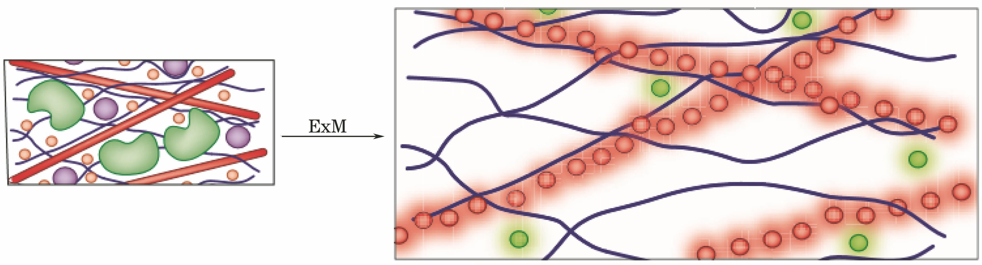
    [15] Chang J B, Chen F, Yoon Y G et al. Iterative expansion microscopy[J]. Nature Methods, 14, 593-599(2017).

    [16] Wei F F, Liu Z W. Plasmonic structured illumination microscopy[J]. Nano Letters, 10, 2531-2536(2010).

    [17] Fleischmann M, Hendra P J, Mcquillan A J. Raman spectra of pyridine adsorbed at a silver electrode[J]. Chemical Physics Letters, 26, 163-166(1974). http://www.sciencedirect.com/science/article/pii/0009261474853881

    [18] Wei F F, Lu D, Shen H et al. Wide field super-resolution surface imaging through plasmonic structured illumination microscopy[J]. Nano Letters, 14, 4634-4639(2014). http://pubs.acs.org/doi/pdf/10.1021/nl501695c

    [19] Grandin H M, Stadler B, Textor M et al. Waveguide excitation fluorescence microscopy: A new tool for sensing and imaging the biointerface[J]. Biosens Bioelectron, 21, 1476-1482(2006). http://www.ncbi.nlm.nih.gov/pubmed/16137877

    [20] Diekmann R, Helle Ø I, Øie C I et al. Chip-based wide field-of-view nanoscopy[J]. Nature Photonics, 11, 322-328(2017). http://www.nature.com/abstractpagefinder/10.1038/nphoton.2017.55

    [21] Betzig E. Proposed method for molecular optical imaging[J]. Optics Letters, 20, 237-239(1995). http://europepmc.org/abstract/MED/19859146

    [22] Hafi N. Grunwald M, van den Heuvel L S, et al. Fluorescence nanoscopy by polarization modulation and polarization angle narrowing[J]. Nature Methods, 11, 579-584(2014).

    [23] Frahm L, Keller J. Polarization modulation adds little additional information to super-resolution fluorescence microscopy[J]. Nature Methods, 13, 7-8(2015). http://www.ncbi.nlm.nih.gov/pubmed/26716557

    [24] Hafi N. Grunwald M, van den Heuvel L S, et al. Reply to "Polarization modulation adds little additional information to super-resolution fluorescence microscopy"[J]. Nature Methods, 13, 8-9(2015).

    [25] Strack R. Imaging: Orientation mapping in super-resolution[J]. Nature Methods, 13, 902(2016). http://www.nature.com/nmeth/journal/v13/n11/full/nmeth.4061.html

    [26] Zhanghao K, Chen L, Yang X S et al. Super-resolution dipole orientation mapping via polarization demodulation[J]. Light: Science & Applications, 5, e16166(2016). http://www.nature.com/uidfinder/10.1038/lsa.2016.166

    [27] Cruz C A V, Shaban H A, Kress A et al. . Quantitative nanoscale imaging of orientational order in biological filaments by polarized superresolution microscopy[J]. Proceedings of the National Academy of Sciences of the United Stated of America, 113, E820-E828(2016). http://www.ncbi.nlm.nih.gov/pubmed/26831082