• Laser & Optoelectronics Progress
  • Vol. 56, Issue 10, 101001 (2019)
Jinghui Chu, Xiaochuan Li, Jiaqi Zhang, and Wei Lü*
Author Affiliations
  • School of Electrical and Information Engineering, Tianjin University, Tianjin 300072, China
  • show less
    DOI: 10.3788/LOP56.101001 Cite this Article Set citation alerts
    Jinghui Chu, Xiaochuan Li, Jiaqi Zhang, Wei Lü. Fine-Granted Segmentation Method for Three-Dimensional Brain Tumors Using Cascaded Convolutional Network[J]. Laser & Optoelectronics Progress, 2019, 56(10): 101001 Copy Citation Text show less
    Position relationship amongwhole tumor, tumor core and enhancing tumor
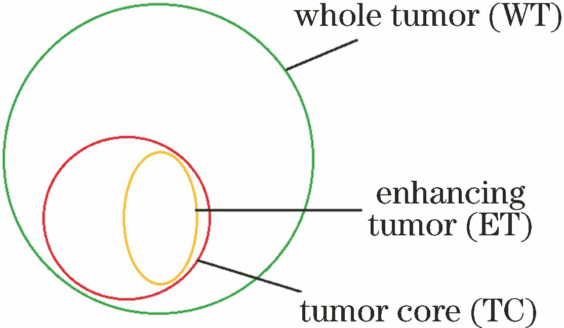
    Fig. 1. Position relationship amongwhole tumor, tumor core and enhancing tumor
    Segmentation system based on cascaded 2.5D convolutional neural network
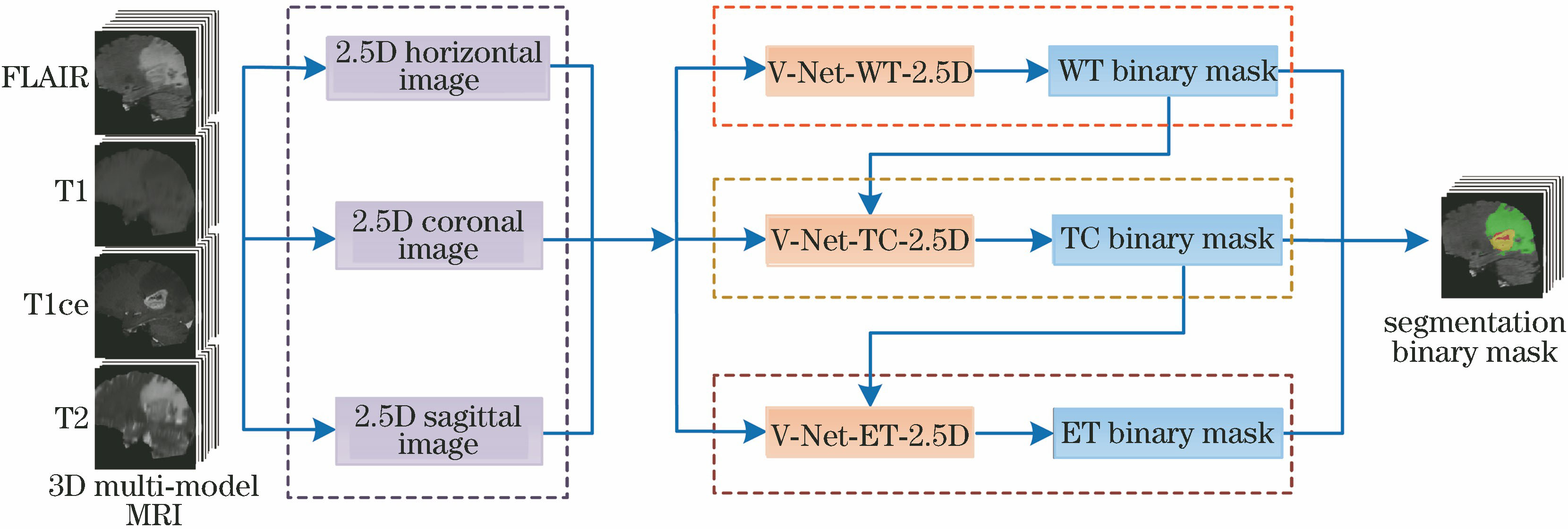
    Fig. 2. Segmentation system based on cascaded 2.5D convolutional neural network
    Structural diagram of proposed 2.5D V-Net
    Fig. 3. Structural diagram of proposed 2.5D V-Net
    Structural diagram of whole tumor segmentation module
    Fig. 4. Structural diagram of whole tumor segmentation module
    Structural diagram of tumor core segmentation module
    Fig. 5. Structural diagram of tumor core segmentation module
    Structural diagram of enhancing tumor segmentation module
    Fig. 6. Structural diagram of enhancing tumor segmentation module
    MRI images in three sections for different modes. (a) T1 images; (b) T2 images; (c) FLAIR images; (d) T1ce images; (e) label mask for fine segmentation
    Fig. 7. MRI images in three sections for different modes. (a) T1 images; (b) T2 images; (c) FLAIR images; (d) T1ce images; (e) label mask for fine segmentation
    Segmentation results of different networks in three directions for sample 1. (a) T1ce images; (b) end-to-end V-Net; (c) cascaded V-Net-3D; (d) cascaded V-Net-2D; (e) cascaded V-Net-2.5D
    Fig. 8. Segmentation results of different networks in three directions for sample 1. (a) T1ce images; (b) end-to-end V-Net; (c) cascaded V-Net-3D; (d) cascaded V-Net-2D; (e) cascaded V-Net-2.5D
    Segmentation results of different networks in three directions for sample 2. (a) T1ce images; (b) end-to-end V-Net; (c) cascaded V-Net-3D;(d) cascaded V-Net-2D; (e) cascaded V-Net-2.5D
    Fig. 9. Segmentation results of different networks in three directions for sample 2. (a) T1ce images; (b) end-to-end V-Net; (c) cascaded V-Net-3D;(d) cascaded V-Net-2D; (e) cascaded V-Net-2.5D
    Qualitative comparison of segmentation results before and after fusion. (a) FLAIR images; (b) T1ce images; (c) horizontal prediction; (d) coronal prediction; (e) sagittal prediction; (f) fused prediction
    Fig. 10. Qualitative comparison of segmentation results before and after fusion. (a) FLAIR images; (b) T1ce images; (c) horizontal prediction; (d) coronal prediction; (e) sagittal prediction; (f) fused prediction
    MethodEnd-to-end-V-NetCascaded V-Net-3DCascaded V-Net-2DCascaded V-Net-2.5D
    Dice WT0.87140.88320.89860.9071
    Dice TC0.75620.79240.84130.8542
    Dice ET0.65240.71220.79190.8140
    Table 1. Comparison of various algorithms for ten-fold cross-validation
    DirectionHorizontal predictionCoronal predictionSagittal predictionFused prediction
    Dice WT0.89890.88080.89120.9071
    Dice TC0.84570.83670.84010.8542
    Dice ET0.80810.79220.80010.8140
    Table 2. Segmentation performance comparison of 2.5D V-Net fed by images in different directions
    LossfunctionCrossentropyDice lossDice loss+Jaccard loss
    Dice WT0.90420.90510.9071
    Dice TC0.84110.85010.8542
    Dice ET0.79080.80890.8140
    Table 3. Segmentation performance comparison based on different loss functions
    Jinghui Chu, Xiaochuan Li, Jiaqi Zhang, Wei Lü. Fine-Granted Segmentation Method for Three-Dimensional Brain Tumors Using Cascaded Convolutional Network[J]. Laser & Optoelectronics Progress, 2019, 56(10): 101001
    Download Citation