
Yanqi Chen, Jiurun Chen, Zhiping Wang, Yuting Gao, Yonghong He, Yishi Shi, An Pan, "Fast full-color pathological imaging using Fourier ptychographic microscopy via closed-form model-based colorization," Adv. Photon. Nexus 4, 026001 (2025)
- Advanced Photonics Nexus
- Vol. 4, Issue 2, 026001 (2025)
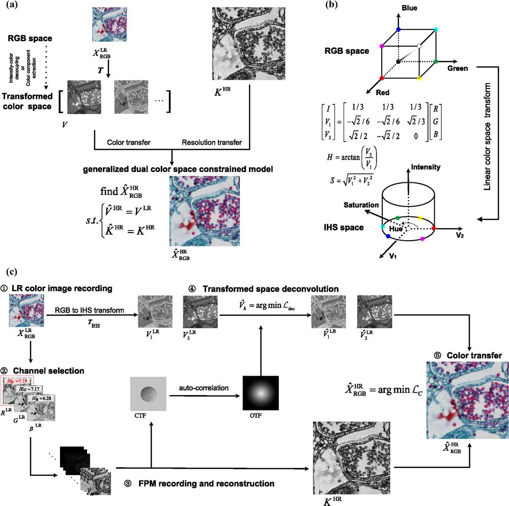
Fig. 1. Principle of gCFPM. (a) Schematic of generalized dual-color-space-constrained model for color transfer; (b) principle of RGB-to-IHS transform; and (c) flow chart of gCFPM using IHS-RGB space constraints.

Fig. 2. Full-color imaging via gCFPM. (a) Imaging result of the sample; (b) photograph of cat stomach smooth muscle section; (c) enlarged view of ROI; and (d) full process time comparison of 3-FPM (three-channel FPM) and gCFPM.
Fig. 3. (a) Photograph of stratified epithelium section; (b) photograph of lycopodium sporophyll spike longitudinal section; (c) imaging result of (a); and (d) imaging result of (b).
Fig. 4. Comparison of different colorization methods. (a) Simulation result; (b) RMSE and CSSIM comparison in (a); and (c) comparison using experimental data. The upper row in (c) shows the result of ROI 3 in the stratified epithelium section; the lower row in (c) shows the result of ROI 1 in lycopodium sporophyll spike L.S.
Fig. 5. (a) Imaging results of gCFPM and 20× objective with defocus ranges from
|
Table 1. Quantitative comparison of different methods in experiments.
|
Table 2. Assessment of gCFPM based on the extended linear transform.


Set citation alerts for the article
Please enter your email address

© Copyright 2018-2021 | Chinese Laser Press. All Rights Reserved 沪ICP备15018463号-20